Subacromial Impingement Syndrome - Podcast Version
TeachMeSurgery
Subacromial impingement syndrome (SAIS) refers to inflammation and irritation of the rotator cuff tendons as they pass through the subacromial space, resulting in pain, weakness, and reduced range of motion within the shoulder.
SAIS encompasses a range of pathology including rotator cuff tendinosis, subacromial bursitis, and calcific tendinitis. All these conditions result in inflammation between the coracoacromial arch and the supraspinatus tendon or subacromial bursa.
It occurs most commonly in patients under 25 years, typically in active individuals or in manual professions, and accounts for around 60% of all shoulder pain presentations, making it the most common pathology of the shoulder.
The Subacromial Space
The subacromial space lies below the coracoacromial arch and above the humeral head and greater tuberosity of the humerus. The coracoacromial arch consists (lateral to medial) of the acromion, the coracoacromial ligament (anterior to the acromioclavicular joint), and coracoid process.
Within the subacromial space run the rotator cuff tendons, the long head of biceps tendon, and the coraco-acromial ligament, all surrounded by the subacromial bursa which helps to reduce friction between these structures.
By TeachMeSeries Ltd (2020)
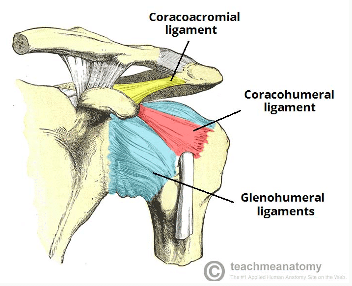
Figure 1
The ligaments of the shoulder joint
Pathophysiology
The underlying cause of subacromial impingement syndrome can be divided into intrinsic and extrinsic mechanisms
Intrinsic mechanisms involve pathologies of the rotator cuff tendons due to tension, which include:
- Muscular weakness – weakness in the rotator cuff muscles can lead to muscular imbalances resulting in the humerus shifting proximally towards the body
- Overuse of the shoulder – repetitive microtrauma can result in soft tissue inflammation of the rotator cuff tendons and the subacromial bursa, leading to friction between the tendons and the coracoacromial arch
- Degenerative tendinopathy – degenerative changes of the acromion can lead to tearing of the rotator cuff, which allows for proximal migration of the humeral head
Extrinsic mechanisms involve pathologies of the rotator cuff tendons due to external compression, such as:
- Anatomical factors – Congenital or acquired anatomical variations in the shape and gradient of the acromion
- Scapular musculature – Reduction in function of the scapular muscles, that normally allow the humerus to move past the acromion on overhead extension, may result in a reduction in the size of the subacromial space
- Glenohumeral instability – Any abnormality of the glenohumeral joint or weakness in the rotator cuff muscles can lead to superior subluxation of the humerus, causing an increased contact between the acromion and subacromial tissues
Clinical Features
The most common symptom of SAIS is progressive pain in the antero-superior shoulder region. The pain is classically exacerbated by abduction in the affected shoulder and relieved by rest, and may be associated with weakness and stiffness secondary to the pain.
Two common examination signs can be elicited in cases of SAIS (specifically for subacromial impingement):
- Neer’s Impingement test – The arm is placed by the patient’s side, fully internally rotated and then passively flexed, and is positive if there is pain in the anterolateral aspect of the shoulder
- Hawkin’s test – The shoulder and elbow are flexed to 90 degrees, with the examiner then stablising the humerus and passively internally rotates the arm, and the test is positive if pain is in the anterolateral aspect of the shoulder
Differential Diagnoses
- Muscular tear (e.g. rotator cuff tear, long head of biceps tear) – the weakness will persist despite the shoulder pain being relieved
- Neurological pain (e.g. thoracic outlet syndrome, cervical radiculopathy, brachial plexus injury) – any weakness will likely be associated with paraesthesia or pain, yet the weakness will persist despite the shoulder pain being relieved
- Frozen shoulder syndrome (adhesive capsulitis, calcific tendinitis) – stiffness will persist even if the pain is relieved
- Acromioclavicular pathology (e.g. acromioclavicular arthritis, glenohumeral arthritis) – presents with a more generalised pain, also with weakness and stiffness related to pain
Investigations
The diagnosis of impingement is a clinical one, however it is often confirmed via additional imaging.
MRI imaging of the affected shoulder is often the mainstay of imaging for SAIS. Features that can be seen in affected individuals include formation of subacromial osteophytes and sclerosis, subacromial bursitis, humeral cystic changes, and narrowing of the subacromial space.
Adapted from RSatUSZ (Own work) [CC BY-SA 3.0 (https://creativecommons.org/licenses/by-sa/3.0)], via Wikimedia Commons

Figure 2
MRI scan showing subacromial impingement with a partial rupture of the supraspinatus tendon
Management
Conservative management is the mainstay of treatment in most cases. Patients should have sufficient analgesia, typically non-steroidal inflammatory drugs, and regular physiotherapy.
For those who require further intervention, corticosteroid injections in the subacromial space can be trialled. Patients should be educated appropriately with adequate warm-up techniques prior to exercise and monitoring for early signs of worsening impingement.
Surgical Intervention
If SAIS persists beyond 6 months without response to conservative management, surgical intervention is recommended.
Surgical intervention is particularly useful in patients with a reduced range of movement. Current surgical techniques include:
- Surgical repair of muscular tears, most commonly the supraspinatus and long head of biceps tendon, resulting in an improvement in range of motion
- Surgical removal of the subacromial bursa, a bursectomy, increasing the subacromial space and reducing pain
- Surgical removal of a section of the acromion, an acriomioplasty, increasing the subacromial space and reducing pain
However, recent evidence from a randomised surgical trial showed that surgical decompression appeared to offer no extra benefit over arthroscopy alone, and indeed that the benefits seen from the surgical intervention might only be the result of a placebo effect or the increased post-operative physiotherapy.
Complications
Complications of SAIS include rotator cuff degeneration and tear, adhesive capsulitis, cuff tear arthropathy and complex regional pain syndrome.
If diagnosed and assessed early, SAIS resolves with conservative management in 60 – 90% of patients.
Key Points
- Subacromial impingement syndrome refers to the inflammation and irritation of the rotator cuff tendons as they pass through the subacromial space
- Presents with progressive pain in the anterior superior shoulder, typically worsening by abduction and relieved by rest
- Diagnosis is a clinical one however MRI imaging can be useful to confirm the diagnosis and assess for further complications of the condition
- Mainstay of management is conservative, with limited evidence advocating surgical intervention